Viruses
| Microbes | Virus examples | Virus discussion |
Reference
Enger & Ross
Audesirk & Audesirk
Ch 19
Virus Wiki
| HyperPhysics***** Biology | R Nave |
Viruses
|
Index Reference Enger & Ross Audesirk & Audesirk Ch 19 Virus Wiki | |||
|
Go Back |
Comparative Sizes of Microbes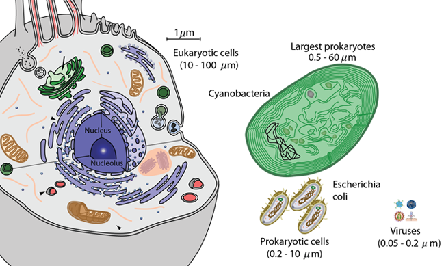
|
Index Reference Audesirk & Audesirk Ch 19 | ||
|
Go Back |
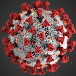
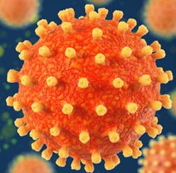
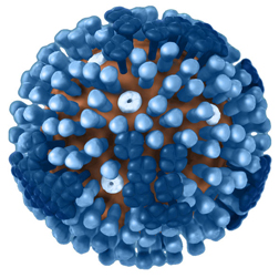
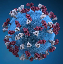
Virus ExamplesViruses occur in a vast array of shapes and configurations. These images provide some information about the shape and structure of the viruses, but the size of their features is generally below the resolution of light microscopes, being in the range of 0.05 - 0.2 μm or 50 - 200 nanometers compared to the range of visible light wavelengths 400-750nm. While the colors used to display the structures are useful for visualization, they are not to be taken literally because the entire virus is smaller than the wavelength of those colors of light. The structural details can be visualized with electron microscope images since the effective electron wavelengths can be made small enough to resolve the details by using high energy electrons.
|
Index Reference Enger & Ross Audesirk & Audesirk Ch 19 Karp Ch. 1 | |||||||||||||||
|
Go Back |
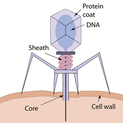
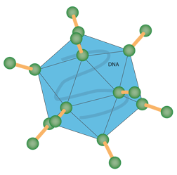
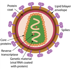
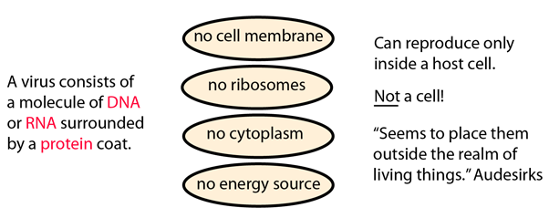
The Varied World of VirusesKarp (the main source of this material) describes viruses as "obligatory intracellular parasites" which cannot reproduce unless they are inside a host cell where they "hijack" its resources. Outside of a living cell, the virus exists as a particle called a virion, which is little more than a molecular package. Nevertheless, there is a tremendous variety of such packages. They contain genetic material which may be single-stranded or double-stranded and composed of either DNA or RNA. Some viruses have as few as three or four different genes, while others have as many as several hundred. The genetic material is surrounded by a protein capsule, or capsid. Considering their enormous impact on human life, it is remarkable to note that virions are just molecular aggregates, inanimate particles that by themselves are unable to reproduce, metabolize, or carry out any of the other activities associated with life! Yet each type of virion has on its surface a protein that is able to bind to a particular surface component of its host cell. The size of viruses is so small that it is less than the wavelength of visible light, so they cannot be seen with a light microscope, but they can be "tagged" with a fluorescent molecule, enabling them to be caused to glow when excited by UV light and be tracked by a light microscope. By this technique it was demonstrated that virus particles could penetrate a host cell wall in less than a tenth of a second and move to the cell's nucleus within 15 minutes! Once there, they can take over the cell's manufacturing capacity to induce it to produce components for the virus. All this for a virion which appears as an inanimate particle! Karp describes viral infections as mostly occurring in two basic types: 1. The virus arrests the normal synthetic activities of the host cell and redirects the cell to use its available materials and energy to manufacture specific viral nucleic acids and proteins. Unlike the process of cell reproduction, these components directly assemble themselves into new virions. Ultimately the cell ruptures or "lyses" to release a new generation of viral particles. 2. The infecting virus does not lead to the death of the host cell, but instead inserts (integrates) its DNA into the DNA of the host cell. The integrated viral DNA is called a provirus. An integrated provirus can have different effects depending upon the type of virus and host cell.
|
Index Reference Enger & Ross Karp Ch. 1 | |||
|
Go Back |