Arterial Ultrasound Scan
High frequency ultrasound in the 7-12 MHz region is used for high resolution imaging of arteries which lie close to the surface of the body, such as the carotid arteries. Using a nominal sound velocity of 1540 m/s in tissue, the sound wavelength in tissue for a 7 MHz sound wave can be obtained from the wave relationship v = fλ.
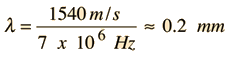 |
Using the general principle of imaging that you can't see anything smaller than the wavelength suggests a 0.2 mm ultimate resolution limit.
In addition to imaging the arterial walls, the ultrasound techniques can measure the blood flow velocity by making use of the Doppler effect. The reflected ultrasound is shifted in frequency from the frequency of the source, and that difference in frequency can be accurately measured by detecting the beat frequency between the incident and reflected waves. The beat frequency is directly proportional to the velocity of flow, so continuous recording of the beat frequencies from the different parts of the the arteriy gives you an image of the velocity profile of the blood flow as a function of time.

The sketches above are conceptual only; no attempt has been made to accurately scale velocities and colors. But hopefully it illustrates the use of false color imaging to give an instant view of the distribution of velocities present. The lower part of the illustration contains an intensity modulated power spectrum in which the beat frequency and therefore the speed of the blood is on the vertical axis. Such spectra are produced by analysis of the reflected ultrasound using a mathematical process called a fast Fourier transform (FFT) in which the distribution of reflected power as a function of frequency is extracted. This is done repetitively and the results plotted as a function of time (horizontal axis). The distance vertically from the axis indicates the beat frequency and therefore the velocity of flow. The relative amount of power reflected at a given velocity value is indicated by the brightness of the display at that point. A single uniform flow speed would give a single bright line, so the display indicates a considerable range of velocities present in the flow at any time. Note that at the time of the peaks, essentially all of the blood has a fairly high velocity since the part of the spectrum near the horizontal axis is dark.
 |
Carotid ImageThis example of a clinical carotid image is taken during the relatively quiet period between the peaks. Note that red indicates a slower flow than blue, but in the same direction since carotid flow does not reverse. So the implication of red and blue is not the same as in the astrophysical red shift of light from stars. Note the background grayscale image which is formed from the pulse-echo ranging data. |
Taken literally, the above image would suggest a fairly uniform flow velocity over the cross-section of the artery. But that is not the nature of the expected laminar flow, in which there is expected to be a velocity profile with the highest velocity on the center line and dropping toward zero velocity at the walls. I'm guessing that the red range of the false color image is set to include a wide range of low velocities.
A higher ultrasound frequency like 12 MHz gives a shorter wavelength and therefore higher resolution, but that advantage is partially canceled by the fact that the higher frequency is attenuated more in tissue. So judgments must be made about the relative merits of deeper penetration (low frequency) vs higher resolution (higher frequency).
The ultrasound sources are generally tuned ceramic wafers of a material such as PZT which are driven by applying an AC voltage at the design frequency. The voltage causes mechanical vibration by the piezoelectric effect.
The ultrasound scans can detect the buildup of plaque in the arteries. Besides the direct imaging of the narrowed vessel, the Doppler information can be converted into false color images which profile the flow velocity. Flow in a region of obstruction must be at a higher velocity to maintain the flowrate, and that velocity information is confirmation of a narrowing of the vessel.
| Ultrasound | Doppler pulse probe |
| Speed of Sound in Tissue |
Traveling wave concepts
| HyperPhysics***** Sound | R Nave |