Rods and Cones

The retina contains two types of photoreceptors, rods and cones. The rods are more numerous, some 120 million, and are more sensitive than the cones. However, they are not sensitive to color. The 6 to 7 million cones provide the eye's color sensitivity and they are much more concentrated in the central yellow spot known as the macula. In the center of that region is the " fovea centralis ", a 0.3 mm diameter rod-free area with very thin, densely packed cones.

The experimental evidence suggests that among the cones there are three different types of color reception. Response curves for the three types of cones have been determined. Since the perception of color depends on the firing of these three types of nerve cells, it follows that visible color can be mapped in terms of three numbers called tristimulus values. Color perception has been successfully modeled in terms of tristimulus values and mapped on the CIE chromaticity diagram.

Rod and cone distribution More cone details More rod details
Index

Vision concepts

Color vision concepts

Reference

Williamson & Cummins
Ch 6,10
 
HyperPhysics***** Light and Vision R Nave
Go Back


Rod and Cone Density on Retina

Cones are concentrated in the fovea centralis. Rods are absent there but dense elsewhere.

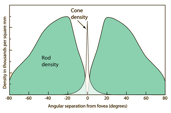
Measured density curves for the rods and cones on the retina show an enormous density of cones in the fovea centralis. To them is attributed both color vision and the highest visual acuity. Visual examination of small detail involves focusing light from that detail onto the fovea centralis. On the other hand, the rods are absent from the fovea. At a few degrees away from it their density rises to a high value and spreads over a large area of the retina. These rods are responsible for night vision, our most sensitive motion detection, and our peripheral vision.

The above illustration does make it appear that there are no cones outside the fovea centralis, but that is not true. The blue cones in particular do extend out beyond the fovea.

Cone details Rod details

Reference:

Anatomical distribution of rods and cones

Physiological principles for effective use of color

Rods and cones

Index

Vision concepts

References

Williamson & Cummins
 
HyperPhysics***** Light and Vision R Nave
Go Back


Cone Details

Current understanding is that the 6 to 7 million cones can be divided into "red" cones (64%), "green" cones (32%), and "blue" cones (2%) based on measured response curves. They provide the eye's color sensitivity. The green and red cones are concentrated in the fovea centralis . The "blue" cones have the highest sensitivity and are mostly found outside the fovea, leading to some distinctions in the eye's blue perception.

The cones are less sensitive to light than the rods, as shown a typical day-night comparison. The daylight vision (cone vision) adapts much more rapidly to changing light levels, adjusting to a change like coming indoors out of sunlight in a few seconds. Like all neurons, the cones fire to produce an electrical impulse on the nerve fiber and then must reset to fire again. The light adaption is thought to occur by adjusting this reset time.

The cones are responsible for all high resolution vision. The eye moves continually to keep the light from the object of interest falling on the fovea centralis where the bulk of the cones reside.

Rod and cone discussionRod and cone distribution
Index

Vision concepts

Reference

Williamson & Cummins
Ch 6,10
 
HyperPhysics***** Light and Vision R Nave
Go Back


"Blue" Cone Distinctions

The "blue" cones are identified by the peak of their light response curve at about 445 nm. They are unique among the cones in that they constitute only about 2% of the total number and are found outside the fovea centralis where the green and red cones are concentrated. Although they are much more light sensitive than the green and red cones, it is not enough to overcome their disadvantage in numbers. However, the blue sensitivity of our final visual perception is comparable to that of red and green, suggesting that there is a somewhat selective "blue amplifier" somewhere in the visual processing in the brain.

The visual perception of intensely blue objects is less distinct than the perception of objects of red and green. This reduced acuity is attributed to two effects. First, the blue cones are outside the fovea, where the close-packed cones give the greatest resolution. All of our most distinct vision comes from focusing the light on the fovea. Second, the refractive index for blue light is enough different from red and green that when they are in focus, the blue is slightly out of focus (chromatic aberration). For an "off the wall" example of this defocusing effect on blue light, try viewing a hologram with a mercury vapor lamp. You will get three images with the dominant green, orange and blue lines of mercury, but the blue image looks less focused than the other two.

Index

Vision concepts

Reference

Williamson & Cummins
Ch 6,10
 
HyperPhysics***** Light and Vision R Nave
Go Back


Rod Details

The rods are the most numerous of the photoreceptors, some 120 million, and are the more sensitive than the cones. However, they are not sensitive to color. They are responsible for our dark-adapted, or scotopic, vision. The rods are incredibly efficient photoreceptors. More than one thousand times as sensitive as the cones, they can reportedly be triggered by individual photons under optimal conditions. The optimum dark-adapted vision is obtained only after a considerable period of darkness, say 30 minutes or longer, because the rod adaption process is much slower than that of the cones.

The rod sensitivity is shifted toward shorter wavelengths compared to daylight vision, accounting for the growing apparent brightness of green leaves in twilight.

While the visual acuity or visual resolution is much better with the cones, the rods are better motion sensors. Since the rods predominate in the peripheral vision, that peripheral vision is more light sensitive, enabling you to see dimmer objects in your peripheral vision. If you see a dim star in your peripheral vision, it may disappear when you look at it directly since you are then moving the image onto the cone-rich fovea region which is less light sensitive. You can detect motion better with your peripheral vision, since it is primarily rod vision.

The rods employ a sensitive photopigment called rhodopsin.

Rod and cone discussionRod and cone distribution
Index

Vision concepts

Reference

Williamson & Cummins
Ch 6,10
 
HyperPhysics***** Light and Vision R Nave
Go Back


Rods Do Not See Red!

The light response of the rods peaks sharply in the blue; they respond very little to red light. This leads to some interesting phenomena:

Red rose at twilight: In bright light, the color-sensitive cones are predominant and we see a brilliant red rose with somewhat more subdued green leaves. But at twilight, the less-sensitive cones begin to shut down for the night, and most of the vision comes from the rods. The rods pick up the green from the leaves much more strongly than the red from the petals, so the green leaves become brighter than the red petals!

The ship captain has red instrument lights. Since the rods do not respond to red, the captain can gain full dark-adapted vision with the rods with which to watch for icebergs and other obstacles outside. It would be undesirable to examine anything with white light even for a moment, because the attainment of optimum night-vision may take up to a half-hour. Red lights do not spoil it.

These phenomena arise from the nature of the rod-dominated dark-adapted vision, called scotopic vision.

Index

Vision concepts

Reference

Williamson & Cummins
Ch 6,10
 
HyperPhysics***** Light and Vision R Nave
Go Back